Conjoined Sternophagus Twin Monster: A Cause of Dystocia in Murrah Graded Buffalo- Juniper Publishers
Journal of Dairy & Veterinary Sciences- Juniper Publishers
Abstract
A 6 years old murrah graded buffalo in second
lactation with the history of 305 days of gestation was presented in the
jurisdiction of veterinary hospital Hamirpur, Himachal Pradesh. The
animal had started showing signs of labor 3 hours prior to its
presentation. Per vaginal examination revealed fetus in anterior
presentation, dorso-sacral position with two heads and four forelimbs,
which confirmed it to be a case of fetal dystocia due to either twin
pregnancy or fetal abnormalities. Per vaginal manipulation of fetus was
attempted by mutations but proportionately large size of abnormal fetus
impeded the process. Adopting caesarian section as means of fetal
delivery was opted and a sternophagus twin monster was delivered.
Keywords: Sternophagus; Murrah graded buffalo; Dystocia; Caesarian section
Introduction
Dystocia is a most commonly observed in bovines and
the condition is developed when the birth process is hampered by some
physical obstacle or functional defects [1]. Any fetal defect such as
fetal monster may result in distortion of body configuration and can
become a reason of dystocia in bovines [2,3]. An incidence of fetal
monstrosities was recorded up to 7.9% [4] to 12.8% [5] in river
buffaloes. Most of the monstrosities observed in water buffalo and
little data is available for swamp buffaloes. Monstrosities causing
dystocia are most likely to be relieved by caesarian section [3,6,7]. It
is strenuous for the fetal monster to pass through the birth canal
either owing to the altered shape they possess or their relative size.
Most usual encountered fetal monsters are Schistosoma reflexus,
Perosomus elumbis, conjoined twins and cyclopia [8].
The conjoined twins are two fetuses secured together
and arise typically from a single ovum and are monozygotic occurring
because of incomplete division of a fertilized ovum and display great
discrepancy from incomplete duplication to almost complete separation of
two individuals, joined in just a few places [8]. Conjoined twins are
non-inherited teratogenic defects [2]. It is believed that some factors
are responsible for the failure of twins to separate after the 13th day
after fertilization those results in conjoined twins [9]. Conjoined
twins monozygotic in origin may be fused medially at different parts of
body but the cranial fusion is noted to be commonest of all [8].
Case History
A Murrah graded buffalo aged about 6 years in second
parity was presented with the history of 305 days of pregnancy was
presented in the jurisdiction of veterinary hospital Hamirpur. Animal
was straining for last 3 hours with two visible limbs in the vagina.
When the animal was presented, the water bags had already ruptured as
the case was manipulation by local para-veterinary staff. The animal was
alert and was in good body condition with rectal temperature of 100.9º
F. Vaginal mucus membrane was congested and edematous with negligible
lubrication. Per vaginal examination revealed complete dilation of
cervix with more than two fore limbs and two heads in the birth canal
suggestive of the conjoined twin fetus. Both the fetuses were in
anterior longitudinal presentation with dorso-sacral position. Per
vaginal delivery was attempted to relieve dystocia by mutation, forced
extraction after achieving adequate lubrication of birth canal, yet
unsuccessful to deliver the fetus. Disproportionate size of the mother
and the monster fetus justified caesarian to alleviate the condition.
Result and Discussion
Cesarean was conducted under local anesthesia which
was achieved by using 90ml, 2% lignocaine hydrochloride in linear
infiltration. Caesarian section was performed in the left lateral
recumbancy. An oblique vento-lateral incision was given, and a dead male
conjoined twin monster was removed. The surgical wound was sutured in
routine fashion by using chromic catgut no. 2. The uterus was sutured by
Cushing suturing pattern. The first and second muscle layers were
sutured by lock stitch and simple continuous suture pattern,
respectively. Skin was sutured with silk by using simple interrupted
suture pattern. The buffalo was treated with inj. Strepto-penicillin
5.0gm and inj. Meloxicam 15ml through intramuscular route for 5 days.
The supportive therapy was done with inj. Normal saline and Ringer’s
lactate 4 liter each through intravenous route only once. Antiseptic
dressing of
surgical wound was done with povidone iodine and removal of
suture was done after 12 days of the surgery.
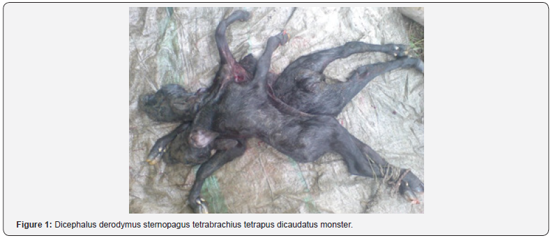
The fetus was conjoined at sternum region of the thorax and
both the heads were opposite to each other. Grossly the fetus
possessed two normal heads, with separate nostrils, eyes and
ears. According to Camon [10] dicephalic fetus could manifest
any of the following feature; Atlodymus (two complete and
separate skulls and single neck), Iniodymus (two skulls with
fusion at the occipital region) and derodymus (two complete
and separate skulls with two separate necks). In accordance with
this nomenclature, the present fetus was derodymus dicephalic,
distomus, tetraopthalmus, tetraotus, tetrabrachius, tetrapus, and
dicaudatus conjoined sternopagus twin monster male buffalo calf
(Figure 1). Similar findings in a buffalo conjoined monster were
reported by Shukla [2].

Conclusion
A rare case of conjoint twin monster in buffalo manifesting
duplication of external body parts was delivered successfully by
cesarean section.
To know more about journal of veterinary science impact factor: https://juniperpublishers.com/jdvs/index.php
To know more about Open Access journals Publishers: Juniper Publishers


Comments
Post a Comment