Post-Partum Uterine Eversion and its Management in A Doe: A Case Report- Juniper Publishers
Abstract
A case of uterine prolapse in 3 years old
non-descript doe is reported. Assessment of prolapsed mass was done
under epidural anesthesia. Uterus was cleaned with potassium
permanganate solution and Lignocaine jelly and Soframycin was applied on
the mass. Oxytocin and broad-spectrum antibiotics were administered
intramuscularly in recommended doses. Aim of present study was to
highlight the management of uterine prolapse in doe.
Keywords: Doe; Epidural anesthesia; Lignocaine; Prolapsed mass
Introduction
Uterine prolapse is an eversion of the uterus which
turns inside out as it passes through the vagina generally occurs
immediately after or few hours of parturition when the cervix is open
and lacks tone [1]. Most common post-partum complication observed in
cows and ewes and less commonly in does [2]. Uterine prolapse in goats
may be complete with both the horns protruding out from vulva or may be
limited to the uterine body [3]. Etiology of uterine prolapse is
unknown, but many factors have been associated with it [1,4]. These
includes poor uterine tone, increased straining caused by pain or
discomfort after parturition, by excessive traction at assisted
parturition or by the weight of retained fetal membranes, conditions
such as tympany and excessive estrogen content in feed leads to
increased intra-abdominal pressure leading to uterine prolapse. Prolapse
that occur more than 24 hours post-partum is extremely rare and is
complicated by partial closure of the cervix, making replacement
difficult or even impossible [5]. Immediately after prolapsed occurs the
tissues appear almost normal, but with passage of time becomes enlarged
and edematous. Some animals may develop hypovolaemic shock secondary to
internal blood loss, laceration of the prolapsed organ or incarceration
of abdominal viscera [6]. Success of treatment depends on the type of
case, duration of case, degree of damage and contamination. Present
study envisages the management of uterine prolapse in goat.
Case History
An adult non-descript doe was presented to the
Teaching Veterinary Clinical complex Palampur, India with complaint
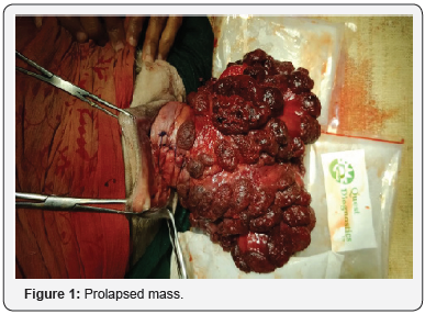
of hanging mass from vulva (Figure 1). Owner reported that doe delivered
two female kids at full term of gestation and subsequently about 3hour
uterine prolapse occurred. At the time of observation, the animal was
dull and depressed. The clinical examination of the animal showed pale
visible mucus membranes, body temperature 990F, heart rate 128/min and
respiratory rate 22/min. Clinico-gynaecological examination revealed,
edematous everted uterine mass soiled with dirt, feces and straw.

Result and Discussion
The goat was administered 2ml of 2% Lignocaine
hydrochloride (LOX®, Neon Labs, India) at first coccygeal space to
attain epidural anesthesia. Prolapsed uterus was gently
washed and disinfected with potassium permanganate solution
(1:1000 dilution) followed by application of icepacks to reduce
edema and volume of the mass. Cervical lacerations were
sutured using simple interrupted suture (Chromic catgut no.2).
Prolapsed mass was washed with cold water and lubricated with
Lignocaine jelly (Lignox®, Neon Labs, India) and Soframycin
ointment (Soframycin®, Sanofi, India). By applying adequate
palm pressure, prolapsed mass was pushed inside the vulva
slowly with alternate pushing of upper and lower surfaces and
simultaneously elevating the hindquarters of the animal.
Further, pressure was applied to push the organ forward to
abdominal cavity ensuring the proper repositioning of the uterus
into its original position. No vulval retention suture was applied.
The animal was administered Inj. Calcium borogluconate (100
ml, slow i/v), Inj. DNS (400 ml, i/v), Inj Haemaccel (100ml,
i/v), Ceftriaxone (200 mg, i/m), Inj. Oxytocin (10 I.U, i/m), Inj
Texableed (5ml, i/m), Inj. Flunixin meglumine (1.5ml, i/m),
Inj. Belamyl (2ml, i.m). Antibiotics and Anti-inflammatory was
continued for three days. Haematinic mixture @1.5g once a day
was given orally. The treatment was continued for three days and
doe recovered uneventfully.
Prolapse of uterus normally occurs during third stage of
labor[3] and in small animals, complete prolapse of both the
uterine horns is usual [4]. Prolapse is reported to occur due to
poor body condition, lack of uterine tone, retension of placenta
and irritation in birth canal during parturition [1]. The common
complications of uterine prolapse may be haemorrhages,
shock, septic metritis, suckling problems, infertility and death.
Uterine prolapse is an emergency, which needs immediate
proper treatment, otherwise interference in the blood supply of
prolapsed mass may resulted into edema, cyanosis and later may
develop into gangrene [7]. Sometimes in delayed cases, partial
contraction of cervix interferes with repositioning, resulting in
reoccurrence of prolapse [8].
Prompt treatment of the condition is essential to prevent
toxemia and death of the animal. Possible cause of death of animal
even after treatment could be hypovolaemic shock as a result of
blood loss due to lacerations in prolapsed uterus. Animals with
uterine prolapse treated promptly recovers without complication
while delay in treatment could result in death of the animal in a
matter of hour or so from internal hemorrhage caused by the
weight of the organ which tears the mesovarium and artery
[3]. Since the animals suffering from the uterine prolapse are
hypocalcaemic [5], therefore calcium borogluconate should be
administered. Broad spectrum antibiotics after replacement of
the prolapsed mass prevent secondary bacterial infection [9].
Shock, hemorrhage and thrombo-embolism are potential sequel
of prolonged prolapsed [3]. However, animal can conceive again
without problem if managed properly [10].
Conclusion
Successful management of post-partum uterine prolapse
was done in doe.
To know more about journal of veterinary science impact factor: https://juniperpublishers.com/jdvs/index.php
To know more about Open Access journals Publishers: Juniper Publishers


Comments
Post a Comment