The Use of Cryo dehydratated Animal Anatomical Segments for Veterinary Anatomy Teaching- Juniper Publishers
Journal of Dairy & Veterinary Sciences- Juniper Publishers
Abstract
The veterinary anatomy teaching in the Veterinary
Schools around the world has been supported by diverse kinds of
innovations such as digital resources and applications, being the use of
cadaver's dissections very limited. In the present study, the use of
cryo dehydrated anatomical pieces of musculoskeletal structures from
large and small animals were experimented for five years and the
vantages and advances were computed. The material was prepared using a
fixation of fresh material with 10% formalin followed by dissections and
freezing-unfreezing sequences until completely be dried. Paints were
performed to give a natural appearance. The material produced, including
complete limbs from large and small animals had a good quality and
preservation of the structures such as ligaments, muscle mass, tendons
and apo euroses.
The topographical relationships were perfectly maintained and the pieces
revealed to be a reliable material for the practical of musculoskeletal
anatomy. The method used was easy applied, very cheap, of stress-free
manipulation and storage and due to its high durability reduced the
discharge of biological wastes and chemical products. The students were
very friendly to this kind of material what reflected in high
coefficient of approval during the practical examinations. The
experience of create additional assignment to teaching how to prepare
the material was successful and have been a great integration
opportunity to students from last years of the course work together and
share experiences with the beginners, when they check anatomic contents
considering the applicability in clinic and surgical practices.
Keywords: Veterinary anatomy teaching; Criodehydration technique; Cadaver conservation
Introduction
It is unquestionable that the recognition of gross
anatomical structures and their normal aspect and positioning in healthy
animals is basic in the veterinary formation. Most of Veterinary
Schools around the world have at least two teaching assignatures at the
curricula about gross anatomy. Historically, the experience in this area
counted with the use of cadaver's dissections but this subject has been
much discussed [1,2]
and not more employed in most universities. Although some studies
suggest that dissection, coupled with associated educational activities,
is an effective pedagogical strategy for learning [3,4], nowadays, the use of digital ways to explain this subject is more frequent to describe muscular structures [5],
being the cadaver's use very limited. Unlike osteological material,
that has a permanent character and is available in the laboratories and
museums where is stored with relative easiness, muscular structures are
expensive to preserve. Many techniques are known to keep this kind of
material for long time [6], but most of them implies in high cost of maintenance in suitable tanks. Digital lectures [7] and three-D applications [8]
based on reconstructions of series of drawings are very popular and
helpful among students because the applications allows to put or remove
layer of muscles and they provide basic knowledge about spatial anatomy [9]
but, in most of them the interpolation of data is far of the natural
structure and students have difficulty when in front an anatomical piece
in the laboratory. Synthetic models are also useful, but have an
expressive cost of acquisition and are not so attractive and interesting
to the student as the laboratory practices. The gross anatomical
dissection is a crucial part of the education of veterinarians because
it takes important manual skills, anatomic knowledge as well as an
understanding of spatial relationships of structures and organs of the
body.
Reasons because practices with cadavers are more
scarce includes the low availability of dead animals intended to this
kind of practices once animals are protected by international laws that
forbid the euthanasia for education or investigation studies, the use of
toxic substances to preserve the material, and need of appropriated
facilities to storage the anatomic pieces, mainly treating of large
animals, in which the discharges of biological fluids and of conserving
may a terrible source of environmental contamination.
At South Brazil, many veterinary schools are focused
mainly in farm animals due to the expressive impact of the cattle
farming in the country economy. Other areas of veterinary medicine,
including large animals, are also imperative, such as the equine clinics
and sheep and swine productions. At Federal University of Pelotas,
classes of veterinary anatomy are offered in the first and second
semesters of the curriculum, and composed by a total of 136 hours each
one, with half charge in laboratory practices. The assignment entitled
Anatomy of Domestic Animals I (ADA I) receive annually about 160
academics and has focus in the musculoskeletal structures, being
comparative among domestic species including both, large and small
animals. Parts of bovine carcasses are obtained by donation from
slaughterhouses located near the University, but just corporeal segments
without economic value are available. Other source of biological
material is the Animal Hospital of the university, where the bodies of
dead animals (small and large size) free of infectious and zoonotic
diseases are carried to the Anatomy Sector to classes use. Given it is a
public institution the university has limited financial resources,
including those destined to buy synthetic models to contemplate all
students and, a small number of technicians to support the laboratories.
Considering the high number of students supported and the suitable
quantities of material in the labs, the logistics is compromised due the
space to storage and posterior discharging of cadavers. To become
viable for classes including until 75 students simultaneous at the
laboratory, since the two last decades, we have applied and developed
anatomical techniques10 to solve these problems. This work has as aim to
present these techniques as well as to show how they have worked which
good results in the veterinary teaching.
Material and Methods
Fresh cadavers from equines, bovines and dogs were
used in present study, the carcasses were obtained from animals that
dead from the Animal Hospital from University of Pelotas.
Cryodehydration technique described by previous work [10]
was incremented with paintings and resin layers, being produced
anatomical pieces with superficial and deep muscle dissections. A quick
pass-by-pass is exposed here:
a. Fresh carcasses were used immediately or frozen at -20 °C for further studies;
b. Carcasses with complete integrity (i.e., without
missing parts and without skin without sections) were preferably
perfused with 10% formalin dilution using the external carotid artery
only on one side of the neck. The solution was spread on the carcass
through the vascular system using the pressure produced by raising a
deposit of formalin solution 2.5m from the ground. The expected
positioning of the head and limbs should be provided prior to this step
because the fixed tissues are difficult to move after the process. The
infusion is complete when muscle stiffness is verified and a foamy
discharge is released from nares.
c. When only body parts are available, multifocal
perfusion through muscle tissues and joint cavities is conducted using
manual injection with needles attached to 50ml syringes. The infusion is
complete when, after all the tissues are exposed to the fixative
solution, including the deeper ones. In addition, the material should be
immersed in 10% formalin tanks within the next 48 hours
d. In both cases, the carcasses are covered with plastic material and stored at room temperature (8-220C) during 24 to 48h.
e. Carcasses are washed with tap water and dissected
as a convenience of the proposed class, using traditional anatomic
dissections techniques for musculature visualization.
f. At this stage, the anatomical pieces can be used
in some lab classes, taking account the partial volatilization of the
formalin, with could be irritant to eyes and nose mucosa.
g. Dissected anatomical pieces pass by at least
twenty cryodehydration sequences, including freezing at -200C and
unfreezing at 10 to 220C, air relative humidity around 7080%. After
that, the material is kept out of the freezer until its complete drying,
with is verified when a fine paper towel compressed in the structures
is removed completely dry.
h. A new careful debriding of tissues using the
bistoury to produce scrapping and removal of dry fascia and perimysium
will expose better the muscular fibers. Removals of periosteum in some
parts are also need and will grant a better recognition of the
structures.
i. Finally, structures can be painted with gouache
painting combination colors closer to the natural appearance of a fresh
tissue. Three coatings of gum composed by acetate of polyvinyl are used.
First coating with 50% of dilution in water and the next two using the
pure glum. Drying time is about 12 hours.
Teaching application of the Material
For ten semesters, this kind of material have been
used in the ADA I and was freely available at labs, which were opened at
least 2 hours daily for extra-class study. In some pieces, structures
were permanently labeled. Other kind of gross anatomical pieces included
those fixed in formalin and cryodehydrated metameric sections.
The presence of students in the labs was monitored as
well as the use that they made of the material and the general
conservation of the anatomical pieces. The performance of the students
in subsequent examinations was computed.
Results
Dehydrated anatomical pieces completed about 70% of
the material used in practical classes of veterinary anatomy during the
study. Some students complementary used digital applications but
synthetic models were not available by the university. The material
produced, including complete limbs from large and small animals had a
good quality and conservation of the structures such as ligaments,
muscle mass, tendons and aponeurosis. The topographical relationships
are perfectly maintained and the material revealed to be a reliable
for practical classes of musculoskeletal anatomy. Some regions of
clinical concernment, as ligaments from distal end of equine and bovine
limbs, including all sesamoids suspensory apparatus were very
distinguishable. Thorax and abdomen of dogs also provide excellent
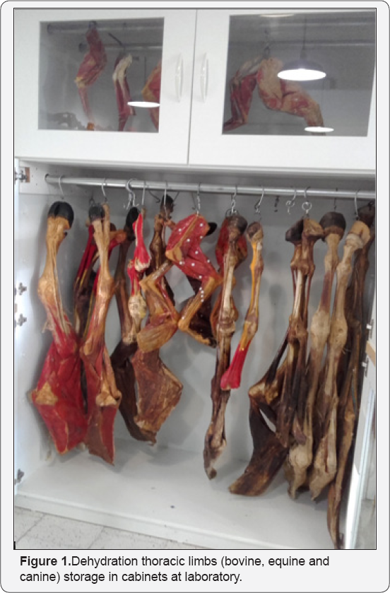
visualization of the muscles. All dehydration pieces were storage in
cabinets with glass door (Figure 1)
and are freely used on laboratory tables by the students. Anatomical
pieces are odourless, dried and resistant to the touch. The students are
friendly to use it because they are very didactic, easy manipulation
and learning. The frequency in the practical laboratory was increased
even when technicians were not available to provide material. Topics
with a good visualization of structures using cryodehidrated anatomical
pieces.
Head of equine and dog
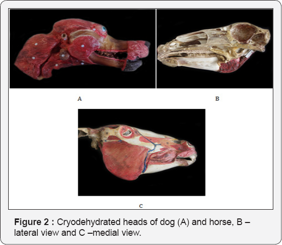
We used to divide using an electric band saw, the
head in left and right antimeric sections, with allowed visualization of
strutures in the medial and lateral aspect. In the medial aspect, it is
appreciable to visualize the encephalon, ethmoid turbinates, nasal
turbinates, hard palate, tongue, pterygoid muscle, parts of hyoid
apparatus, digastric muscle (in horse). From lateral aspect is
visualized most of face musculature, deep or superficial structures
according to the previous dissections (Figure 2).
Limbs of equine, bovine and dog
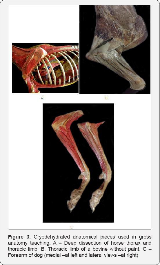
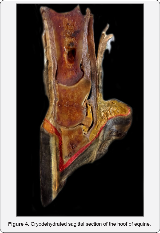
The relationships among muscles and articular structures are clearly verified (Figure 3). Hooves were also sectioned and internal components and their exact positioning and syntopy can be easily studied (Figure 4).
Thorax and abdomen of small animals
These corporal segments were also studied in left and
right antimeric sections and internal organs were removed to allow
highlight to the muscles. Dogs of medium size were chosen and most of
accumulated fat between muscles was manually removed prior dehydration.
In general, material with less than 2 cm of thickness was more quickly
dehydrated. As the most of animals used dead due to traumatic injuries,
the use of hydrogen peroxide 20V, 50% diluted in water was need to
promote cleaning in surfaces with blood impregnation. In practices
classes, the teaching of the muscles of the trunk was also performed
using wet pieces fixed with formalin what allowed the better
understanding the layers of muscles.
Discussion
Cryodehidrated anatomical pieces from the digestive tract have been applied in the some veterinary schools from Brazil [11] with satisfactory results in the anatomical teaching [12].
The methodology explained in the present study for musculoskeletal
structures of large and small animals has been applied with success in
the veterinary anatomy teaching at Federal University of Pelotas for
more than 25 years, but the teaching experience have not evaluated and
published. The great contributions of employing the cryo dehydrated
material as a teaching tool are: the low cost, high durability, easy
handling and storage, low toxicity, long term reduction of environmental
contamination by biological and chemical wastes and the attractive
effect on the students. Similar material is also produced using a
plastination process [13,14]
in which the water and fat are replaced by certain plastics, yielding
specimens that can be touched, do not smell or decay, and even retain
most properties of the original sample [11].
However, this process is not available in most Veterinary Schools
around the world due to high cost of execution, once require specific
equipment and chemical components. The cryo dehydration technique used
in the present work have a low cost, requiring just formalin, domestic
freezer (for small animals) and artisanal material for completion,
easily found in hardware stores. Once prepared, the anatomical material
can be stored for long time (more than 20 years) in places free of
humidity and decomposers insects (coleopterans). This reduces the
logistic and costs to keep large formalin tanks to store body parts. As
water is lost during the process, there is a reduction of approximately
60% of the original weight of the material which became it slighter and
of easy handling [10].
During the practical classes and studies extra-classes, the students
are not exposed to volatile toxic substances becoming this moment more
long and comfortable. Due its great durability, the same piece can be
used for several years without need of replacement, which minimizes the
probability of environment contamination. Lastly, the material produced
in colors or in pale tones (without paints) arouses interest in students
with is expressed in the low disapproval coefficients (8 to 11%)
observed in the practical examinations.
Actually we counted just with a technician to general
activities in the sector that receive also students of zootechny
course, in a total of 600 students annually. Little skilled laborto
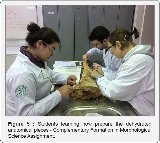
produce the material is required. As the interest of students and
curiosity in to know how to prepare the material have increasing, the
Anatomy Sector decided to create an optional assignment with maximum
10undergraduates to provide specific training (Figure 5).
Two classes are planned each year and, year after year, they have
increasing the anatomical collection available to next students,
sequentially There is a great integration opportunity to students from
last years of the course to work together and share experiences with the
beginners, when they check anatomic contents considering the
applicability in clinics and surgical practices.
Acknowledgment
We are grateful to the students of the Complementary
Formation in Morphological Science Assignment, whose have been
collaborating in preparing anatomical pieces. Thanks go to anonymous
reviewer for the patience to help the English language in this
manuscript.
To know more about our Journal click on Veterinary Journals online free
To know more about open access publishers click on Juniper Publishers



Comments
Post a Comment