Effects of Trypanosomosis on Hemogram and Some Biochemical Parameters of Guinea Pigs Experimentally Infected with Trypanosome Brucei Brucei in Maiduguri, Nigeria- Juniper Publishers
Journal of Dairy & Veterinary Sciences- Juniper Publishers
Abstract
The study was designed to evaluate the effect of
tryponosomosis on Hemogram and some biochemical parameters of guinea
pigs. Guinea pigs of both sexes weighing (5-10kg) were divided into six
groups (A, B, C, D, E and F) with five guinea pigs in each group. At day
zero, to establish the baseline data, all the animals in each of the
six groups were bled for haematology and serum biochemistry and clinical
parameters (rectal temperature, respiratory rate, pulse rate and heart
beats) were recorded while general body condition and physical signs
were also evaluated. Groups A, B and C were intraperitoneally (IP)
inoculated with 1×10⁶ dose of Trypanosoma brucei brucei contained in 0.5
ml of blood. Thereafter, blood samples were collected every other four
(4) days for evaluation of haematology and serum electrolytes through
the experimental period. Group D, E and F was uninfected control. All
the infected groups (A, B, and C) had a pre-patent period of 16 days
with similar levels of parasitaemia of 45.7±3.38 across the groups. The
observed clinical signs among the infected groups (A, B and C) were
pyrexia, pale feet, snout, pinnae and mucous membrane, anaemia,
dullness, emaciation and loss of weight. In group A, a mean parasitaemia
of 2.8 ± 0.84 occurred by day 16 post-infection post infection which
continued to rise significantly without abating (p<0.05) to a peak
count of 120.2 ± 5.48 by day 40 post infection. Similar findings were
noticed across the groups. In groups D, E and F, their respective
pre-infection RBC values of 6.20 ± 1.24, 6.24 ± 1.24 and 6.18 ± 1.24
remained constant (p>0.05).
Abbreviations:
IP: Intraperitoneally; LN: Liquid Nitrogen; NITOR: Nigerian Institute
of Trypanosome and Onchocerciasis; SD: Standard Deviation; PBSG:
Phosphate Buffered Saline Glucose;
Introduction
Trypanosomosis is one of the most important zoonotic
disease commonly found in Africa regions and South America [1]. African
trypanosomosis is caused by a protozoan parasite belonging to the genus
Trypanosoma and is transmitted by tsetse flies to the final host. The
disease is characterized by high morbidity and mortality of infected
livestock. Animal trypanosomosis has been estimated to cost Africa about
US$ 4.5 billion per year [2]. Trypanosomosis affect almost all
vertebrates particularly man and livestock. But wild animals such as
Bovidae and suidae, act as asymptomatic carriers [3]. Biological
transmission (cyclic) of these parasites by tsetse fly (Glossina), is
rampart in tsetse belt zone of Nigeria [3,4]. While arthropod vectors,
haematophagus, of the family Tabanidae, Hippoboscidae, Stomoxynae Haematopota, lyperosia, and Chrysops
species are responsible for the mechanical transmission of the parasite
in the tropics [5,6]. Tsetse flies are efficient transmitters of
trypanosomes especially Trypanosoma vivax which develop in their mouth
[7]. Transplacental transmission of trypanosomes has been reported in
cattle [8]. Trypanosoma vivax, T. congolense and T. brucei have been reported to cause Nagana in cattle, while T. evansi caused surra in camels (Camelus dromedarieus) [9].
Trypanosoma brucei gambiense and T. brucei rhodeseinse are responsible for human sleeping sickness in east and west Africa countries respectively, while T. cruzi
transmitted by triatomid bugs (Triatonia Magista) is responsible for
causing chagas disease in humans in south America [10]. The trypanosomes
group of T. brucei (T. brucei, T. b. gambiense, T. b. rhodensiense and T. evansi) are more of tissue invading (humoral) parasites whereas, T.
congolense, T. vivax and T. cruzi are restricted as hemoparasites
(blood circulation parasites) or (haemic) [11,12]. Animal
trypanosomosis is lethal if left untreated. It causes severe losses
in livestock industries as a result of poor growth, weight loss, and
low milk yield, poor capacity to work, infertility and abortion have
been reported in low levels of infection [3]. Control of the disease
therefore is significant through hematological and biochemical
evaluation that can lead to a reliable diagnosis that can guarantee
optimistic treatment that will restore production in endemic areas
and boost livestock production industries.
Materials and Methods
Source of Trypanosoma Brucei Brucei
The trypanosoma parasites used for the study was “Federe”
strain of Trypanosoma brucei brucei, which was obtained from
NITOR (Nigerian Institute of Trypanosome and Onchocerciasis)
Kaduna State. Nigeria. The organism was isolated from an outbreak
of bovine trypanosomosis in Nassarawa State of Nigeria. It was
identified based on its morphology and negative blood inhibition
and infectivity test and was stabilized by four passage in rats
before storage in liquid nitrogen (LN). Four donor rats were used
to multiply the parasites and transported by road from Kaduna
to the Department of Veterinary Medicine, Faculty of Veterinary
Medicine, University of Maiduguri, Borno State, Nigeria. The
parasites were then maintained in Albino rats by serial passage
until used.
Experimental Animals
Total of Thirty (30) apparently healthy Guinea pigs of both
sexes and of different ages were used in the study. The animals
were purchased from breeders in Plateau State. On arrival, they
were kept in clean and well-ventilated cages in the Large Animal
Veterinary Clinic, Faculty of Veterinary Medicine, university of
Maiduguri, Nigeria. They were routinely screened for ecto, haemo
and endo parasites using standard methods. The Guinea pigs were
housed in a suitable locally made wire mesh cages with sawdust as
bedding, fed with varieties of vegetables and commercial growers
feed (Vital Feeds, PLC, Nigeria), and water provided ad libitum.
They could acclimatize to laboratory condition for two weeks
prior to commencement of the experiment. The experimental
procedure was in accordance with regulations of the Ethical
Committee of the Faculty of Veterinary Medicine, University of
Maiduguri.
Experimental Infection
Four (4) adult albino rats were used as donors of T. brucei
brucei, the rats were purchased from NITOR, Kaduna State.
They were screened for internal and external parasites using
standard method as described by [5]. They were inoculated intra
peritoneally with 0.5 ml of T. brucei brucei parasiteamic blood
with multiple parasites. At 4 days post inoculation, parasiteamia
was established and became obvious. The donor rats were bled via
the tail vein into a petri dish, the blood was diluted with phosphate
buffered saline glucose (PBSG) (pH 7.4). Each Guinea pig in groups
A, B and C were inoculated intraperitoneally with 0.5ml of blood
containing 1.0 × 10⁶ Trypanosoma brucei brucei as quantified
using serial dilution as reported by Herbert & Lumsden [13].
Experimental Design
Thirty Guinea pigs were randomly divided into six groups
with five Guinea pigs in group (A, B, C, D, E and F). At day zero, all
the animals were bled for haematology and serum biochemistry to
establish a baseline data. Thereafter, physical condition and clinical
parameters of all the animals were evaluated (rectal temperature,
respiratory rate, pulse rate and heart beats) and body weights
respectively. Groups A, B and C were inoculated with 0.5ml of blood
containing 1.0 × 10⁶ Trypanosoma brucei brucei intraperitoneally
(IP). Blood sample were collected for haematology and serum
electrolytes profiles at interval of 4 days from day zero until
the end of the experiment. Group A was infected with 0.5 ml of
blood containing 1.0 × 10⁶ Trypanosoma brucei brucei (untreated
control), Group B was infected with 0.5ml of blood containing
1.0 × 10⁶ Trypanosoma brucei brucei (treated with Diaminazene
diaceturate Veriben®) at day 28 post infection (p.i) at the dose
rate of 7.0 mg/kg body weight. Group C was infected with 0.5ml
of blood containing 1.0 × 10⁶ Trypanosoma brucei brucei (treated
with Diaminazene diaceturate (Veriben®) at day 28 post infection
at the dose rate of 3.5mg/kg body weight. While Group D and
Group E were uninfected/untreated (control) and uninfected
treated with diaminazene diaceturate (Verben®) at day 28 (p.i) at
the dose rate of 7.0mg/kg body weight respectively. Group F was
uninfected (treated with diaminazene diaceturate (Veriben®) at
day 28 post infection at the dose rate of 3.5mg/kg body weight.
Post Infection Evaluation of Guinea pigs
All the Guinea pigs were observed daily for the manifestation
of clinical signs of trypanosomosis, which include morbidity and
mortality. Meanwhile, detection of parasitaemia was done every 4
days post day zero and the degree of parasitaemia was projected
by the rapid matching technique as described by [13,14].
Blood Collection
Blood for haematological and serum electrolytes examination
were aseptically collected at 4 days interval preliminary from
day 0 to day 64 post infection. The Guinea pigs were bled using
2ml syringe through cardiac puncture 0.5ml and 1ml of the blood
was transferred into tubes containing anticoagulants (EDTA) and
plain for hematological indices and sera for biochemical profiles
respectively. Hematological and biochemical parameters were
determine as described Henry & Tietz [15].
Statistical Analysis
Data generated were expressed as mean ± standard deviation
(SD) using Two-way analysis of variance was used to compare the
data between groups and value p < 0.05 was considered significant
[16].
Results
The prepatent period in both groups was the same form
4 -
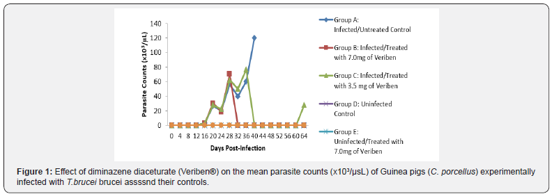
8 days post infection. Following patency, parasitaemia fluctuated
considerably in both groups with a higher recorded and least in
group A and B respectively as presented in Figure 1. There was
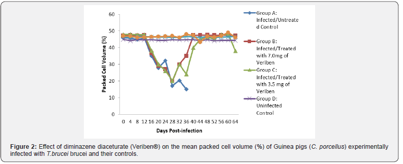
a significant decrease in PCV values of both infected groups
compared with their controls (Figure 2). The decline in PCV
values began with the arrival of trypanosomes in circulation of all
the infected groups with different degrees of anaemia as shown
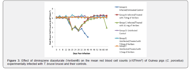
in (Figure 2). The value of RBC declined significantly (p<0.05) in
all the infected groups following parasitaemia without reduction
up to day 40 post infection in group A as presented in (Figure 3).
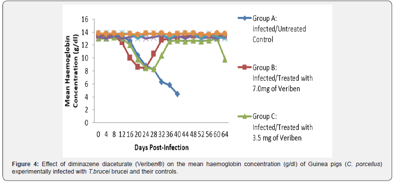
Group A, Hb value significantly decreased following parasitaemia
hence, the Hb value continued to decline significantly (p<0.05)
without abating up to day 40 post infection as documented in
(Figure 4).

From day 16 (pi) when parasitaemia became patent in all
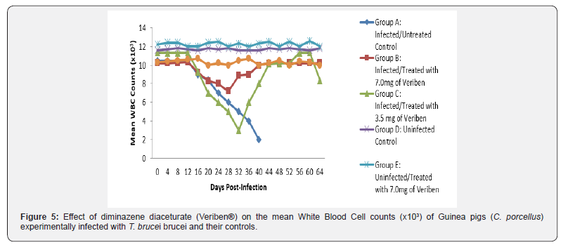
the infected groups, the WBC value continuous to declined
significantly particularly prominent in group A and B as shown
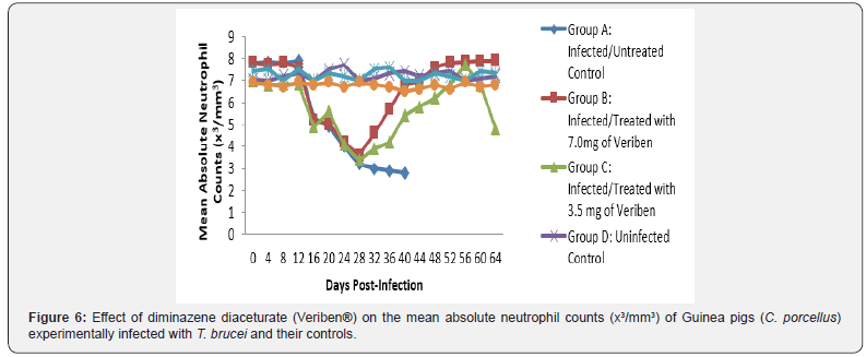
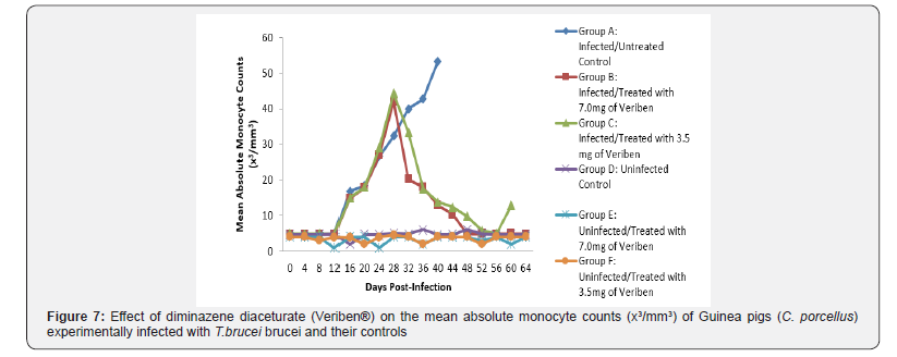
in (Figure 5). The differential leucocytes count of all the infected
groups declined following the establishment of parasitaemia
by day 16 post infection, hence the value continue to decline
significantly (p<0.05) in group A, B and C as indicated in (Figures
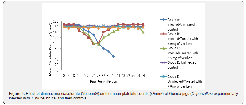
6-8). While there was significant decreased in platelets count in
all the infected groups following parasitaemia appearance in day
16 post infection in group A and B as shown in (Figure 9) In group
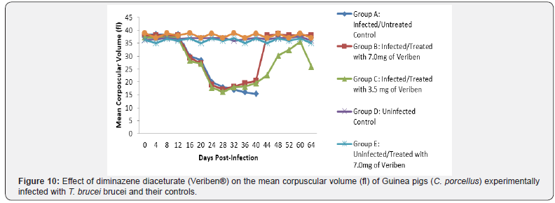
A, the values of MCV decreased significantly (p<0.05) following
establishment of parasitaemia by day 16 post infection similar
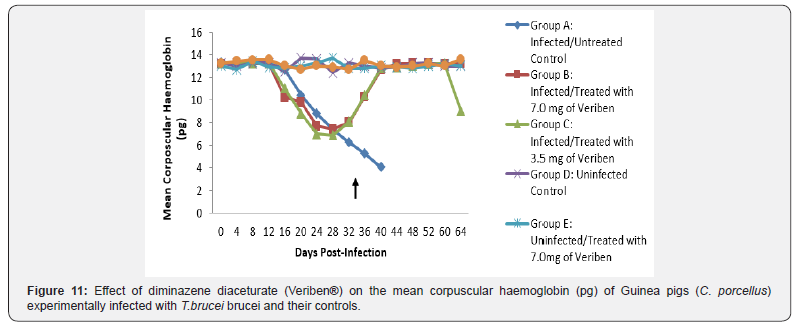
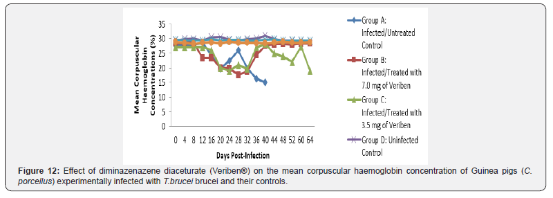
findings was observed on the mean corpuscular haemoglobin
MCH of the Guinea pigs (C. porcellus) as presented in (Figures 10-
12).
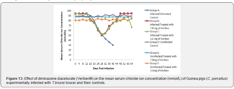
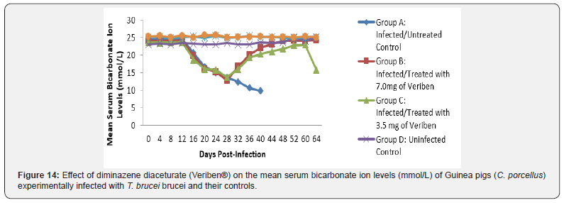
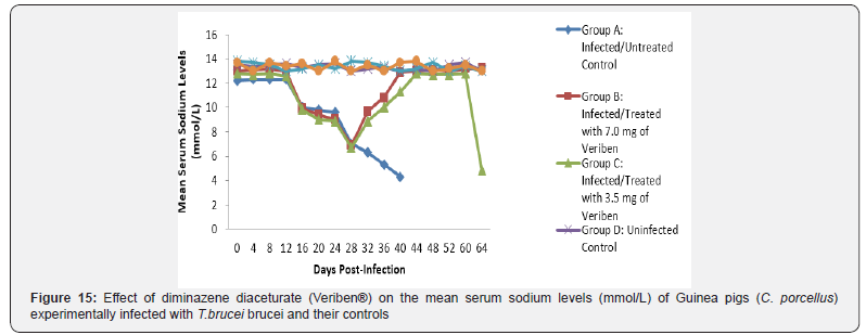
The mean chloride ion concentrations of the Guinea pigs
(C. porcellus) continually decreased following establishment of
parasitaemia by day 16 post infection in all the treated groups
at different days, similar findings was noticed on the mean
bicarbonate ion concentrations of the Guinea pigs (C. porcellus)
at day 16 post infection and mean serum sodium levels of Guinea
pigs (C. porcellus) the serum sodium levels decreased significantly
following establishment of parasitaemia by day 16 post infection
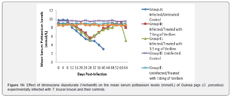
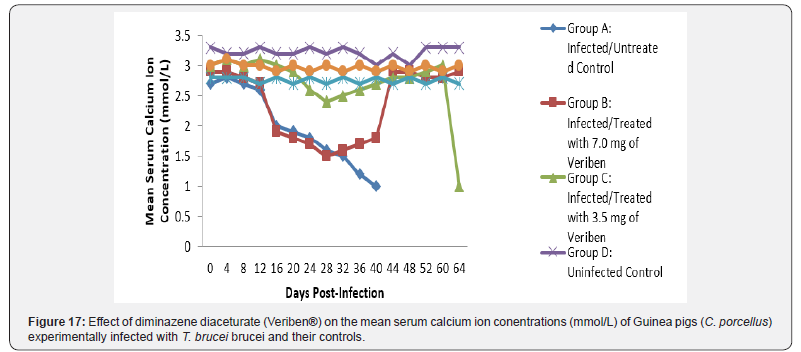
in all the treated groups as show in (Figures 13-15). The mean
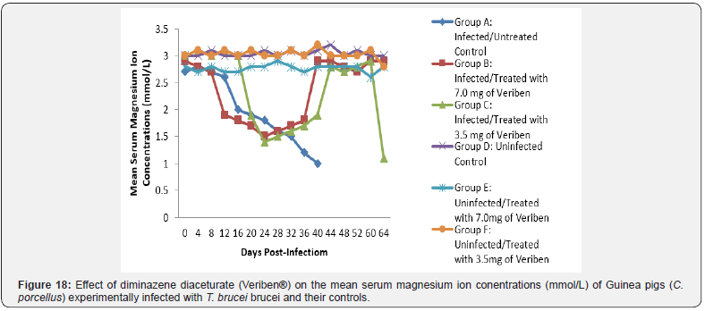
serum calcium ion concentration, mean serum potassium levels
and magnesium ion concentration of Guinea pigs (C.porcellus)
experimentally infected with T.brucei brucei were continually
decreased following establishment of parasitaemia by day 16
post infection in all the treated groups at different days interval as
presented in (Figures 16-18).
Discussion
The main clinical signs observed following experimental
infection were respiratory distress, anemia, raised hair coat,
dullness, anorexia and emaciation, this agreed with findings of
several author reported in several species of laboratory animals
and livestock. After the manifestation of clinical signs, the infected
Guinea pigs shows parasitaemia by day 16 post infection. This is
contrary to the findings of Umar [17] who observed a prepatent
period of 4 days in rats infected with Trypanosoma brucei brucei.
Infected Guinea pigs, started losing weight two weeks post
infection. This may be associated with anorexia observed earlier,
this was like the findings reported by [18,19] in rats infected
with T. brucei brucei and rats infected with T. brucei gambiense
respectively [20] also reported similar findings in rabbits infected
with T. brucei brucei [21].
Despite the sudden changed in red cell parameters by
decreased values of (PCV, RBC, Hb) during bouts of parasitaemia
but gradually increased during the period of low and no
parasitaemia. This tallies with the report of Yusuf [22], who
observed a significant decrease in PCV, Hb, RBC and platelet count
in rats infected with T. b. brucei. This showed an inverse relationship
between parasitaemia and anaemia in most of the infected animals
[23,24]. Anaemia is a consistent feature of trypanosome infections
associated by oxidative stress that inflict damage to erythrocytes
membrane and associate components. Equally, reactive oxygen
radicals generated during trypanosomosis can attack erythrocyte
membranes, which may induce oxidative stress that can trigger
haemolysis [25]. It was also observed in the current study
that only the group of Guinea pigs infected with T. b. brucei and
treated with 3.5mg/kg diaminazene diaceturate (Veriben®)
developed relapse parasitaemia at day 36 post treatment. Relapse
in trypanosomosis has been linked to the parasites moving into
areas of the body that are not accessible to the drugs like the
brain. If infection can continue for some time before treatment
then it is extremely difficult to obtain a permanent cure [26,27].
Also, Waitumbi [26] reported relapse parasitaemia in rabbit
infected with T.brucei brucei following treatment with 25mg/kg of
diaminazene diaceturate. The difference with the current study in
the occurred relapse might be attributed to difference species of
animal used and the drug dosage.
The haematological results of the present study agreed with
earlier studies reported by [28-30]. The low PCV observed in the
infected group may be linked to acute hemolysis that occur due
to the progressive nature of the infection. Previous studies have
shown that infection with trypanosomes resulted in increased
susceptibility of red blood cell membrane to oxidative damage
probably due to depletion or reduction of glutathione on the surface
of the red blood cell [31]. Severity of anaemia has been related to
reflect the intensity and length of parasitaemia. Several reports
[32,33] have also ascribed acute anaemia in trypanosomosis to
proliferating parasites. The low leucocytes (WBC, lymphocytes,
and neutrophils) and platelet counts observed in the infected
group may be attributed to the immunosuppressive actions of
trypanosome infection [34].
The major function of platelets is to activate blood clotting
mechanism and prevent loss of blood. In this study, the low
platelet count observed in the infected untreated groups may
indicate destruction of platelets by toxic products emanating
from the trypanosomes [35]. Low platelet counts may also be
attributed to other factors like pooling of blood into the spleen,
removal of platelets by mononuclear phagocytic system and
increased consumption of platelets by disseminated intravascular
coagulation reaction in trypanosome infection [36]. However,
leukocytosis, lymphopaenia and neutrophilia reported, may
be due to trypanosomosis and these conditions usually occur
as a result of wear and tear syndrome associated with animal
immune system caused by the ever changing in variable surface
glycoprotein of the infecting trypanosomes. The lymphopaenia
encountered among the infected Guinea pigs may probably be due
to an increased demand on the system for lymphocytes, which
is a common requirement in both immune and inflammatory
responses in trypanosomosis [37]. Inefficient recovery of iron
from phagocytized RBCs is known to cause iron deficiency in the
body. Igbokwe & Mbaya [38] reported that dyserythropoiesis
is associated with animal trypanosomosis, and this may be
attributed to the decreased MCV that was observed in the present
study among the infected untreated groups.
The T. brucei brucei infected Guinea pigs were observed to be
associated with marked reduction in serum sodium and chloride
ion levels, and this might have been due to renal tubular damage
of the kidneys. The decreased level of serum potassium observed
in the current study was probably due to dehydration associated
with tissue hypoxia. Reduction in bicarbonate ion (HCO3-) levels
may be probably due to acidosis. The reduction may also be due to
decreased alveolar ventilation and tissues hypoxia similar findings
was reported by [39] in sheep infected by trypanosome.
The low bicarbonate levels can also be attributed to the massive
leakages of some electrolytes from cells and tissues damage.
However, the intermittent increase, low level and subsequent
return of these electrolytes to pre-infection levels suggest the
efficacy of the therapies, otherwise it might be as a result of
massive cell and tissue damage at the terminal phase of this single
infection. The decrease in the levels of calcium that was observed
in this study agrees with the findings reported in cattle infected
with T. congolense [40] and sheep infected with T. brucei brucei.
This is said to be due to the deficiency in the parathyroid hormone
because of the destruction of the parathyroid glands or a decrease
in serum carriers, which in this case happens to be albumin. The
drop in the level of serum magnesium concentration noted among
the T. brucei brucei infected Guinea pigs observed in this study
does not tally with the findings of Sow [41] among donkeys in
Burkina Faso and that of Chaudhary & Iqbal [42] among camels
in Pakistan. This may be as a result of the difference in the species
of animals used. The drop-in magnesium concentration in blood
observed in this study might be due to lowered dietary intake due
to the infections. Biochemical evaluation of the body fluids gives
an indication of the functional state of the various body organs
and biochemical changes in body fluids that result from infections
depend on the species of the parasite and its virulence [43].
Conclusion
It is clearly understood that high dose of Veriben®
administered at the dose rate of 7.0mg/kg and 3.5mg/kg have
the abilities of curbing the state of anaemia, immunosuppression,
and serum electrolytes levels in trypanosome-infected guinea pigs
placed on a dose dependent manner.
To know more about journal of veterinary science impact factor: https://juniperpublishers.com/jdvs/index.php
To know more about Open Access Publishers: Juniper Publishers


Comments
Post a Comment