Report on Isolation and Identification of Brucella Abortus from Aborted Foetus and Lymph Nodes of Beef Cattle in Malaysia- Juniper Publishers
Journal of Dairy & Veterinary Sciences- Juniper Publishers
Abstract
Isolation of living brucellae from tissues or
organs such as aborted foetus and lymph nodes has traditionally provided
the most accurate method for the detection of Brucella infection. The
inoculated medium was incubated at 37 °C for 7 days with 10% CO2.
Bacterial colonies growth on the agar was examined. The bacterial cells
were stained with Gram-staining and Modified Ziehl-Neelsen staining
methods and later were examined under microscope to determine their cell
morphology. Biochemical tests were performed to complete the phenotypic
and metabolic characterisation of the isolates. Brucella abortus were
isolated from stomach content, lung and cotyledon of the aborted foetus;
and supramammary and internal iliac lymph nodes of aborted cattle.
Histopathological examination of lymph nodes found the infiltrations of
microphages, neutrophils and giant cells with vacuolated and engorged
macrophages scattered in the necrotic debris of supramamarry lymph node
of aborted cattle.
Keywords: Brucella abortus; Lymph nodes; Aborted foetus; cattleIntroduction
Brucellosis is an infectious disease caused by Gram
–negative facultative intracellular bacterial organisms of the genus
Brucella. The organisms are pleomorphic, short, slender coccobacilli and
their colonies are small, round, convex, smooth, moist-appearing and
translucent. The organisms are pathogenic for a wide variety of animals
and human beings. Brucella abortus is identified as a major pathogen of
brucellosis in cattle all over the world. The disease usually
asymptomatic in non-pregnant females but adult male cattle may develop
an orchitis. The organisms localise in various lymph nodes of female
cattle such as supramammary, udder, retropharyngeal and mandibular lymph
nodes; internal and external iliac lymph nodes; regional lymph nodes
and uterus. The most consistent lesion of B. abortus infection in cattle
involved the lymph nodes, which were remarkably enlarged and had
follicular hyperplasia with a few giant cells and macrophages Forbes et
al. [1].
In pregnant animals, placentitis will occurred
following infection and abortions usually happened between
the fifth and ninth months of the pregnancy. Although serological tests
are being used for monitoring the herds, bacteriological isolation is
still a gold standard for either screening of the
infection or preparing eradication programs Bricker [2]. Direct and
accurate diagnosis of brucellosis can be made by microbiologic
examination. Currently, identification of B. abortus using conventional
methods is commonly applied in the diagnosis of brucellosis. These
methods provide phenotypic and metabolic characterisations based on
microbiological examinations. Biochemical tests have been used to
distinguish different biotypes of Brucella species. Thus, the objective
of this study was to isolate and identify Brucella abortus from cattle
by using biochemical tests and histopathology examination.
Aborted foetus was obtained from cattle which is
serological positive to brucellosis as detected by CFT. Stomach content,
lung, liver and cotyledon were collected from the aborted foetus. Lymph
nodes namely supramammary, mandibular, suprapharyngeal, mesenteric,
internal iliac and superficial inguinal were collected from cattle that
was slaughtered. Lymph node samples were also collected for histological
examination. All samples were kept in ice-box upon transportation to
the laboratory.
All samples were handle followed the techniques
outline in Manual of diagnostic test and vaccines for terrestrial animal
Nielsen & Ewalt [3]. The samples were inoculated onto Brucellaagar
(Pronadisa, Spain) and McConkey agar (Oxoid, USA). All
specimens were processed inside biosafety cabinet. Inoculated
agar plates were placed in CO2 jar and incubated at 37 °C for 7
days. The growth of Brucella organisms on each agar plate was
observed. Brucellae were stained with modified Ziehl-Neelseen
staining method and their cell morphology was examined under
microscope. Isolated Brucellae were identified and characterized
by standard methods as described by Alton et al. [4]. Isolated
organisms were identified by biochemical tests such as catalase,
oxidase, nitrate reduction, H2S production, urease activity and
growth in the presence of thionin and basic fuchsin.

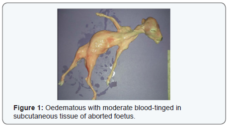
On post-mortem, the aborted foetus were oedematous with
blood-tinged in subcutaneous tissue (Figure 1). The umbilical was
thickened and lungs showed fibrinous pneumonia. The abomasal
contents were turbid, yellow and flaky. Intercotyledonary
placenta was thickened with a yellow gelatinous fluid. The
cotyledons were shown mild to moderate hyperaemic (Figure 2).

Brucella abortus were isolated from stomach content, lung
and cotyledon of the aborted foetus. The organisms were also
isolated from supramammary and internal iliac lymph nodes of
slaughtered cattle that was serologically positive to brucellosis.
The isolates shown typical characteristic of Brucella abortus. The
organisms were grown on Brucella agar but not on McConkey
agar. They required carbon dioxide for their growth and the
colonies were well grown after seven days incubation at 37
°C. The bacterial colonies were raised smooth, dome shape,
white in colour, moist-appearing and transparent when viewed
towards a light source. The isolates were small Gram-negative
coccobacillary cells, present in clumps and stained red by the
modified Ziehl-Neelsen staining method. Biochemical tests
result showed that, the isolates were growth in TSI agar slant and
non-motile. All isolates produced oxidase, catalase and urease.
They also reduced nitrates to nitrites and produced hydrogen
sulphide. The isolates were grown on agar with additional of
basic fuchsin but no growth on agar added with thionin.
The most valuable samples for isolation of Brucellae were
included aborted foetuses (stomach contents, spleen and lung),
fetal membranes, vaginal swabs, milk and semen. In post-mortem,
the suitable samples were lymph nodes such as mammary and
genital lymph nodes; and spleen. The supramammary lymph
nodes were the best source for isolating brucellae in cattle
Sutherland & Searson [5]. Specimens should be collected as
soon as possible following any suspicious abortion or calving.
Specimens should be processed as soon as possible. Ideally,
transportation systems should ensure that specimens arrive
in the laboratory within 24 hours. The procedures used for
collecting and transport of specimens from healthy or clinically
affected animals or post mortem, are critically important for
successful laboratory analyses. These must be done in accordance
with current best practice. Carbon dioxide requirement for
growth, production of hydrogen sulphide and growth in present
of basic fuchsin and thionin were using for confirmation and
differentiate between the Brucella species Nielsen & Ewalt [3].
All isolates were submitted to Veterinary Laboratories Agency
(VLA) United Kingdom for further identification and was
confirmed as Brucella abortus.
To know more about journal of veterinary science impact factor: https://juniperpublishers.com/jdvs/index.php


Comments
Post a Comment