Sarcocystis spp Prevalence in Camel Meat in Jordan-Juniper Publishers
JUNIPER PUBLISHERS-OPEN
ACCESS JOURNAL OF DAIRY & VETERINARY SCIENCES
Keywords
Keywords: Sarcocystosis; Sarcocystis cameli; Camels; Jordan
Introduction
Sarcocystosis caused by Sarcocystis spp is widespread in livestock and has significant economic impact on production of domestic animals as well as public health importance. The parasite has indirect life cycles between two hosts with herbivores as intermediate hosts and carnivores as definitive hosts [1]. The parasite produces tissue cysts in cardiac, striated and smooth muscles of intermediate host. Some Sarcocystis species induce weight loss, general weakness, fever, anorexia, abortion and death in domestic animals [1]. Sarcocystiscameli is the only known species in camels with two different cyst wall: thin-walled and thick-walled cysts. The parasite was first described in camels by Mason [2] in myocardium and esophagus of Egyptian camels. The camels act as intermediate host and becoming infected by ingestion of sporulated oocysts passed in feces of carnivores. Dogs can be infected with S. cameli after ingestion of camel meat, so are important in spreading the infection [3]. Since then the parasite has been reported from almost all of the camel-rearing areas of the world [4]. The parasite has been reported in the United Arab Emirates [5], Egypt [6], Sudan, Iran, Kazakhstan [7], Morocco [8], Saudi Arabia [9], and Iraq [10]. The present paper presents a one year study on the prevalence of sarcosytosis infection in camels in Jordan. Also the study determines distribution patterns of camel Sarcocystis infection in different camel organs.
Material and Method
During a one year period, a total of 97 camels slaughtered at Al- Ramtha slaughterhouse were examined for camel sarcocystosis. The obtained tissue samples for microscopic investigation included diaphragm, heart, esophagus, and skeleton muscles. Collected specimens were divided in to two portions. One portion was kept in 10% formalin for histopathological studies. The second samples was kept in ice-box plastic containers and move to the laboratory for Pepsin-digestion method as described by Dubey et al. and modified by Hamidinejat et al. [11]. The method in summary is done by taking approximately 10g of each tissue organ of the examined camels were crushed and digested for 30min at 40 °C in 50mL of digestion medium containing 1.3g pepsin, 3.5mL HCl, and 2.5g NaCl in 500mL of distilled water. The digested solution was then centrifuged for 5min at 1500RPM. The sediment was smeared on slides, stained by Giemsa stain, and examined at 400X magnification under the light microscope for detection of bradyzoites.
Results

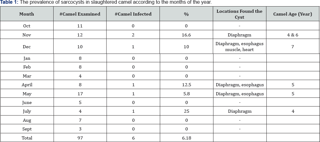
The prevalence of microscopic sarcocysts in slaughtered camel is shown in Table 1. Sarcocystisbradyzoites were found by digestion method in 6out of 97(6.18%) investigated camels at tissue digestion method and 2 out of 97(2.06%) in histopathological methods. Among different organs diaphragm had the highest infection rate (6.18%), followed by the esophagus (3.09%) and heart (1.03%). Also, the infection rate was higher in camels of 4 to 7 years old than younger camels of 1 to 3 years old.
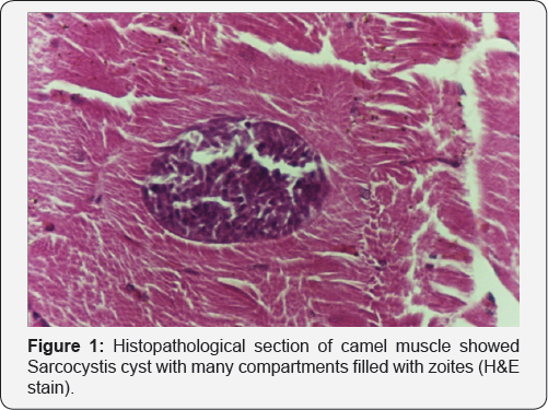
In histopathological sections the sarcocysts were seen embedded between muscle fibers with no inflammation or other evidence of pathological changes around the mature sarcocysts (Figure 1). The morphology of the Sarcosytis reported in this paper of thin wall cyst and most probably Sarcocystiscameli.

Discussion
In this study, the prevalence of sarcocystosisin camels in Jordan is very low (6.6%) comparing to the prevalence of camel sarcocytosisin other countries. The prevalence rate was reported 86.3 % in Iran, 91.6% in Iraq [12], 66.3% in Afghanistan [13], 50% in the United Arab Emirates [14], 88.4% in Saudi Arabia [15]. This may be due management system when stray dogs are under control and not available in the vicinity of camel herds. Analysis of results on distribution of Sarcocystisin this study in different organs showed higher infection rate in diaphragm and then in the esophagus. Fatani et al. [9] reported diaphragm of camels to be the most common site. Other researchers reported higher incidence in esophagus [15,16], while Shekarforoush et al. [14] found heart as the most infected organ. This predilection differences may be due to different S. cameli strains or definite host origin. There is considerable confusion concerning Sarcocystis species in camels. According to Debby et al. two morphologically distinct sarcocysts have been reported in camels; the thin-walled cysts are generally found in diaphragm, heart and esophagus, while the thick-walled cysts are present only in esophagus. By transmission electron microscopy (TEM) two structurally distinct sarcocysts were recognized by unique villar protrusions not found in sarcocysts from any other host. Sarcocysts of S. cameli had villar protrusions of type 9 j. The sarcocyst wall had upright slender villar protrusions, up to 3.0μM long and 0.5μM wide; the total thickness of the sarcocyst wall with ground substance layer was 3.5μM. On each villar protrusions, there were rows of knob-like protrusions that appeared to be interconnected. The villar protrusions had microtubules that originated at midpoint of the ground substance and continued up to the tip; microtubules were smooth, without any granules or dense areas. Bradyzoites were approximately 14-15*3-4μM in size with typical organelles. Sarcocystisip penisarcocysts had type 32 sarcocyst wall characterized by conical villar protrusions with an electron dense knob. The total thickness of the sarcocyst wall (from the base of ground substance to villar protrusions tip) was 2.3-3.0μM. The villar protrusions were up to 1.2μM wide at the base and 0.25μM at the tip. Microtubules in villar protrusions originated at midpoint of ground substance and continued up to tip; microtubules were crisscrossed, smooth and without granules or dense areas [15-18].
In the present study infection rates risk in higher aged camels was observed [19-26]. Our result is in accordance with Woldemeskel & Gumi [12] on Ethiopian camels. Higher chance of acquiring infection in older animals has been reported by Shekarforoush et al. [14] in camels.
For more Open Access Journals in Juniper Publishers please click on: https://juniperpublishers.com/open-access.php
For more articles in Open Access Journal of Dairy & Veterinary sciences please click on: https://juniperpublishers.com/jdvs/index.php


Comments
Post a Comment